
In this article:
- What is the MPA screening process?
- Why does the MPA use flow cytometry for detection?
- Assay Setup optimizes screening conditions
- What data does an MPA screen return?
- How do we know a given screen performed as it should?
- How do we decide what counts as a hit?
- How does Validation confirm or rule out hits?
- Appropriate controls distinguish between constant- and variable-region binding
- How a clean result adds to the weight of evidence for your molecule
- A validated off-target identifies a protein, not the risk
This article is part 9 of a series about specificity testing. Be sure to read part 7, about how we designed a protein library that represents the human membrane proteome.
Summary
Before a biotherapeutic enters the clinic, its developers need to know if it binds any unexpected off-targets. The Membrane Proteome Array (MPA) screens a molecule against roughly 6,000 human membrane proteins to answer that question. This article describes how a test article moves from initial setup through validated, quantified results, the advantages of flow cytometry detection, and why a result showing no off-target binding is a finding in its own right. It includes insights into how we designed an assay that achieves sensitive detection while minimizing false positives.
What is the MPA Screening Process?
The Membrane Proteome Array (MPA) is a cell-based protein array that individually expresses roughly 6,000 human membrane proteins. It gathers quantitative binding data for every protein in the library, showing which proteins a molecule binds and which it does not.
While this article focuses on the MPA, pairing it with screening on our Secreted Proteome Library provides a more complete picture of the interactions a molecule has with the proteins it is likely to encounter in vivo. Learn more in Beyond the Membrane: Secreted Proteins and Comprehensive Specificity Testing.
An MPA study runs in four steps:
Assay Setup. We establish optimal binding conditions for the test article, including the cell line and the screening concentration.
MPA Library Screen. We screen the test article against the full membrane protein library. Proteins whose signal meets the criteria for follow-up are called hits.
Validation Assay. Each hit is retested individually in a titration experiment to confirm genuine off-target binding.
Enhanced Binding Analysis. Currently offered as a follow-on service, this step further characterizes a validated off-target to inform risk assessment.
Each step builds on the last, moving from broad, high-sensitivity detection toward increasingly specific characterization. This article covers setup, screening, and validation—and why you can trust the results. It draws on information provided to the FDA as part of the ISTAND qualification process.
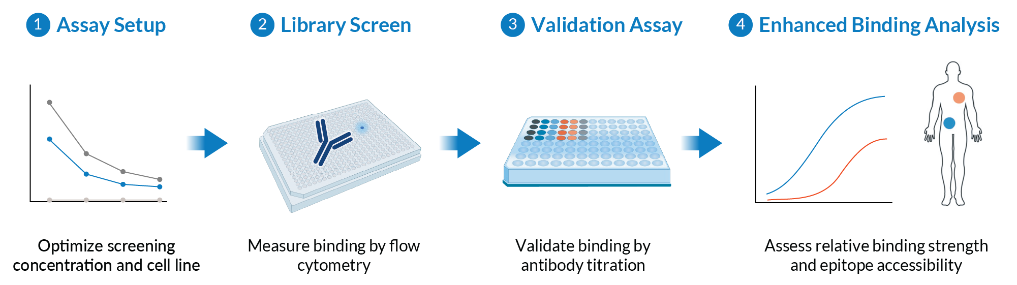
An MPA study runs in sequential steps, moving from broad detection toward increasingly specific characterization. An optional fourth step, an Enhanced Binding Analysis study, further investigates any validated off-target interactions.
Why does the MPA use flow cytometry for detection?
All Membrane Proteome Array (MPA) assays use high-throughput flow cytometry to detect test-article binding, via fluorescently labeled secondary antibodies. Flow cytometry is a well-established method in antibody discovery and characterization, and all instruments undergo routine preventive maintenance and daily calibration by qualified personnel under our ISO 9001–certified quality processes.
Four properties make flow cytometry especially well-suited to specificity testing.
Native cellular proteins. Flow cytometry works on whole cells. Proteins are presented in their native conformations without fixatives, similar to how they appear in vivo.
Selective analysis of cell populations. Because flow cytometry measures multiple cellular parameters and cell markers simultaneously, we can define which cells to include in a measurement.
Quantitative measurement. MFI is a continuous numerical readout, which makes rigorous statistical analysis possible. And because flow cytometry reads individual cells, each sample yields hundreds to thousands of measurements rather than one aggregate reading. This granularity provides statistical power that is not achievable through immunohistochemistry- or immunofluorescence-based assays.
Well-established controls. Appropriate controls rule out artifacts at every step. A positive control confirms that the test article is being detected at all. For antibody formats, we use cells expressing membrane-tethered Protein A, which the antibody binds through its Fc region. Cells transfected with empty plasmid give a direct read on background.
Because binding is measured the same way at every stage, results stay comparable from one step to the next, and from one study to another.
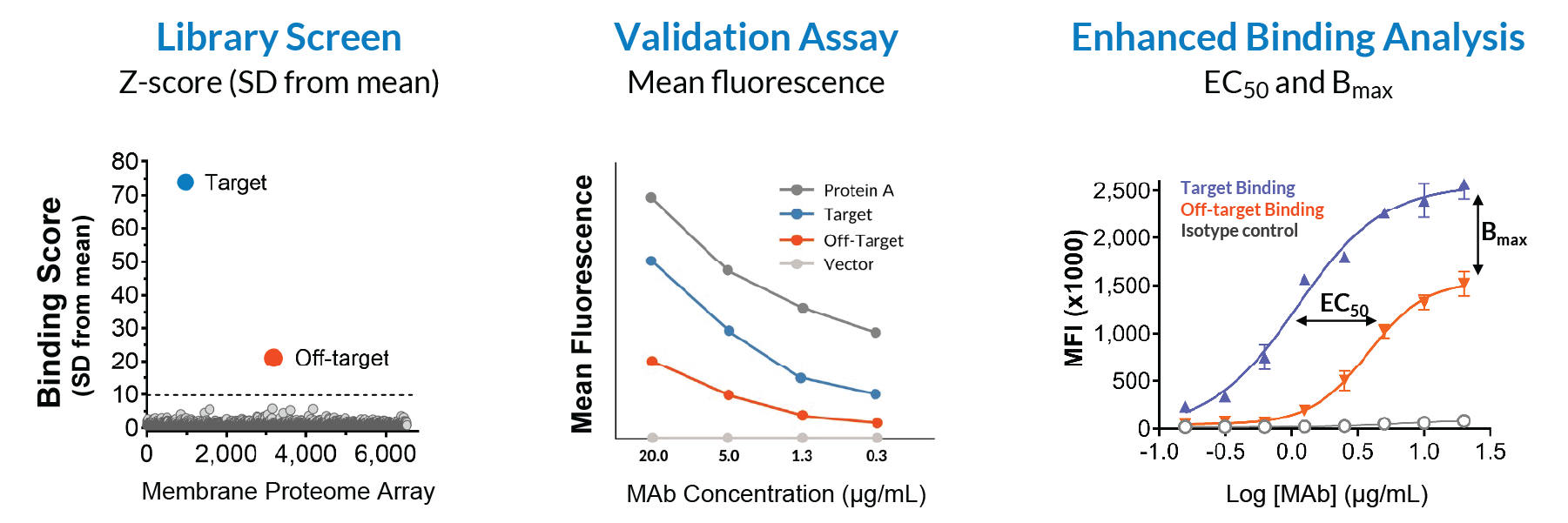
Flow cytometry provides quantitative outputs for each experiment in an MPA study. Results are generated based on Mean Fluorescence Intensity (MFI) signals for binding strength, which provide quantitative and reproducible binding measurements.
Assay Setup optimizes screening conditions
Every MPA study begins with Assay Setup to confirm detection and establish the optimal screening parameters.
Detection. We confirm that the appropriate secondary detection method works for the test article before proceeding to the screening step.
Cell line. Human HEK-293T cells are preferred for screening, because they support native protein folding and appropriate post-translational modifications. However, if HEK-293T cells endogenously express the intended target, background can be high enough to obscure real binding. In that case, we screen instead on avian QT6 cells, which have been validated for high expression levels and native conformations while minimizing interference from endogenous target expression.
Screening concentration. We measure background binding against cells transfected with empty plasmid, define what counts as high background for that molecule, then screen at the highest concentration that stays within those limits—typically 5–20 µg/mL for a monoclonal antibody. Screening at high concentration maximizes the chance of detecting real off-target interactions.
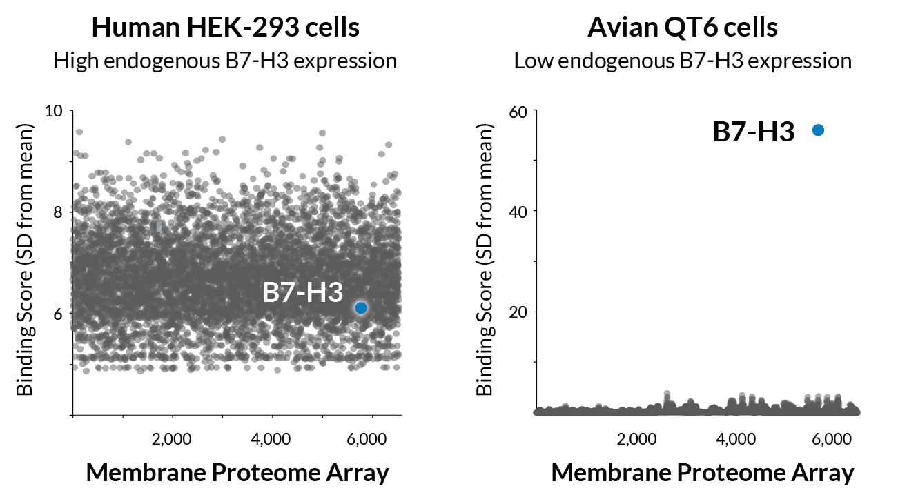
The immune checkpoint protein B7-H3 (CD276) is highly expressed in HEK-293T cells, producing high background binding in an MPA screen. Avian QT6 cells produce a clean result.
What data does an MPA screen return?
A Membrane Proteome Array (MPA) screen returns a binding measurement for every protein in the library, recorded as mean fluorescence intensity (MFI). Most of those measurements fall within the background range.
To make measurements comparable across proteins and between studies, each protein’s binding is expressed as a Binding Score: a z-score in units of standard deviation from the mean signal across the library. This is a quantitative measure, and it lets us compare relative binding across proteins.
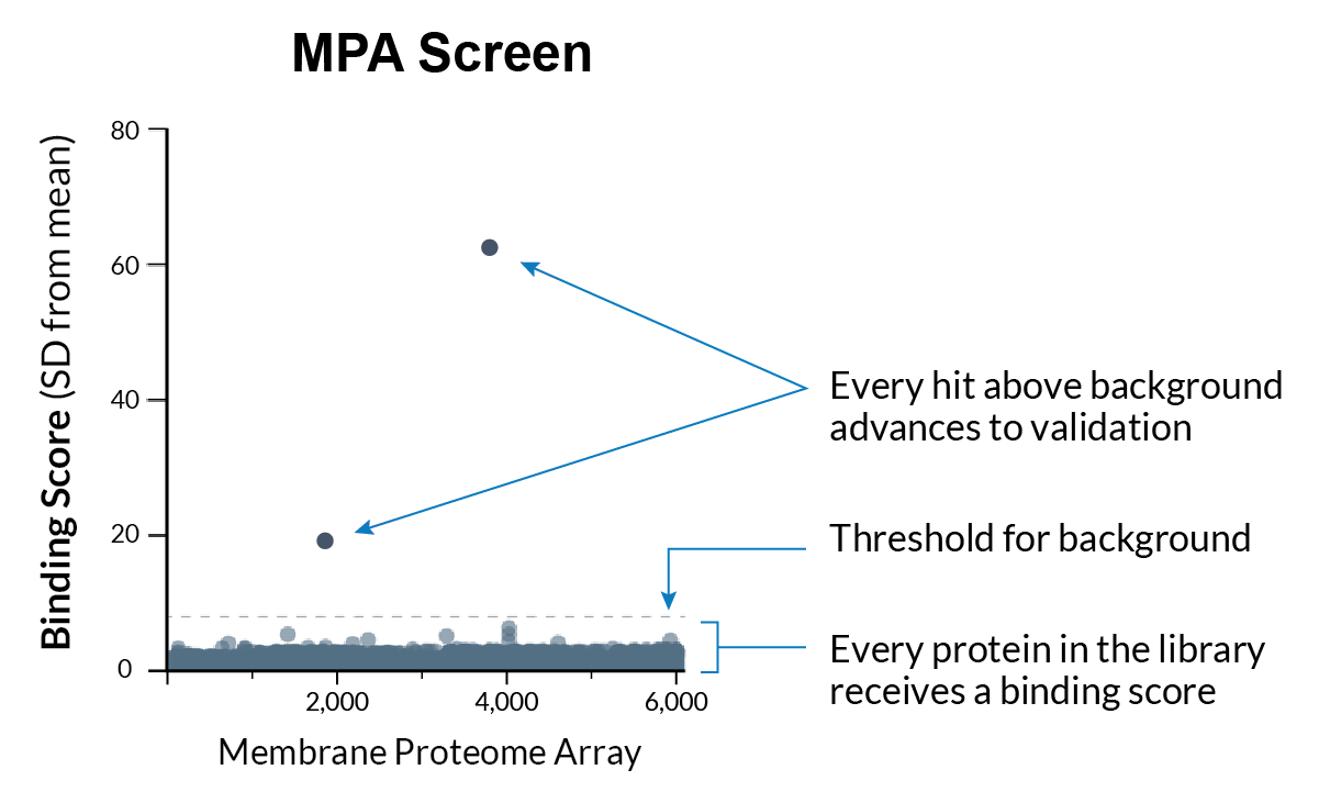
An MPA screen returns a Binding Score for every protein in the library, expressed in units of standard deviation from the mean. Proteins meeting the criteria for follow-up are called as hits.
How do we know a given screen performed as it should?
A clear sign that the screen performed as it should is detection of the molecule’s intended target, which we can confirm if we know its identity. Additional controls and monitoring steps back it up.
Alongside the detection controls described earlier, an internal control protein is tested on every Membrane Proteome Array (MPA) plate in every experiment, confirming that the proteome expressed consistently after transfection.
Every screen also receives an Assay Performance Score, a measure of how cleanly binding signal separates from background across the run. The score tells us how much confidence to place in a particular screen.
Monitoring the Assay Performance Score over time reveals any departure from historical trends as it happens, allowing us to immediately investigate the cause. A score outside the expected range prompts a re-evaluation of the screen and the analysis. If an operational or a procedural error is found, the test article can be re-screened.
If the score falls below the level at which binding can be reliably distinguished from noise, we do not report the result. These cases are rare, and they usually trace back to sample quality, such as aggregation or impurities. When it happens, we talk with the customer about next steps.
How do we decide what counts as a hit?
The working threshold for calling a hit is three standard deviations above the mean, a conventional cutoff in high-throughput screening (Malo et al., 2006). It rests on an established statistical principle: in a normal distribution, fewer than 0.3% of data points are expected to lie farther than 3SD from the mean. In practice, this threshold captures relevant interactions for follow-up while limiting the rate of false positives. Two practices govern how we apply it.
A protein must clear the threshold in both replicates. Every protein in the library is measured in two independent replicates. Because the replicates are independent, an artifact affecting one measurement will not reproduce in the other.
Proteins that meet these criteria are called hits, and every hit goes on to Validation.
How does Validation confirm or rule out hits?
Because the MPA screen is designed to prioritize sensitivity, we conduct a separate Validation experiment to distinguish genuine off-target binding from artifacts. It runs on every hit, in every project, at every service level.
Each hit is retested against the test article in a four-point dose-response titration, and hits are validated or ruled out based on defined criteria. Validated hits are named in the study report along with their binding data.
The practical effect is that you will never receive a list of unconfirmed hits to chase down on your own.
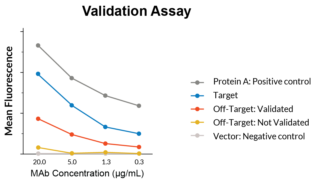
In Validation, each hit is retested in a four-point dose-response titration. Binding that tracks concentration is validated. Binding that does not is ruled out.
Appropriate controls distinguish between constant- and variable-region binding
Off-target binding is not always driven by the molecule’s variable CDR regions. It can also be mediated by the Fc constant region. Since the MPA library includes human Fc receptors, any Fc-containing construct may bind them.
To distinguish between the two, we can run a matched isotype control alongside your molecule in validation studies. Customers can provide their own or choose from our bank of validation control molecules.
Any Fc-mediated binding appears in the study report as a validated interaction, but it is flagged as non-CDR-specific and kept separate from specificity findings that involve the variable domain. Whether Fc receptor binding warrants follow-up depends on the final product. If the final construct retains it, it’s worth investigating.
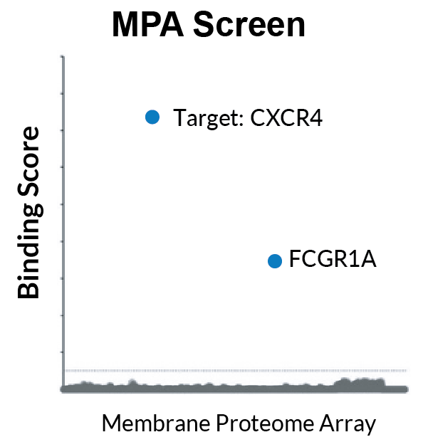
In an MPA screen, an IgG1 format test molecule shows binding to Fc gamma receptor 1A (FCGR1A). Data based on a customer case study.
How a clean result adds to the weight of evidence for your molecule
About two-thirds of the molecules we have screened have no validated off-target binding at all. That is the most common outcome of an MPA study.
A clean finding carries weight because the assay is tuned to find interactions. Proteins are over-expressed so that even low-affinity interactions generate signal, and the test article is screened at a concentration that maximizes sensitivity while minimizing background.
When a screen finds no validated off-target interactions, developers can proceed with confidence, adding to the weight of evidence that a molecule binds only its intended target. And that evidence can be included directly in a regulatory submission.
A validated off-target identifies a protein, not the risk
Together, the MPA Screen and Validation identify precisely which proteins a molecule binds. But they cannot determine whether a given off-target interaction is biologically meaningful. That’s why detecting an off-target is not a signal to abandon a candidate. Rather, it is the starting point for a set of focused, answerable questions that allow for further investigation into any potential safety or functional liabilities.
Looking ahead
The next article covers Enhanced Binding Analysis, the next step if an MPA study identifies a validated off-target. This service further characterizes a named off-target, determining how strongly a test article binds an off-target compared with its intended target, where in the body the protein is expressed, and whether the binding site is likely to be reachable by a circulating drug.