
In this article:
- How was the MPA library designed?
- What’s in the library beyond the canonical membrane proteome?
- How well does the MPA represent human diversity?
This article is part 7 of a series about specificity testing. Be sure to read part 6, about the consequences of off-target binding.
Summary
Specificity testing is ultimately a patient safety question: does this antibody bind only what it’s supposed to? Answering it requires a library that represents the human proteins a biotherapeutic will encounter in vivo, in their biologically relevant conformations. The Membrane Proteome Array (MPA) library was purpose-built to do exactly that. Anchored in FDA guidance and built through deliberate design choices, it captures roughly 6,000 membrane proteins — canonical and non-canonical, sex-balanced, developmentally diverse — representing 94% of the human membrane proteome.
How was the MPA library designed?
Earlier articles in this series established that off-target binding is consequential, common, and frequently missed by traditional specificity testing methods. The Membrane Proteome Array (MPA) was designed to address that gap. Here, we look at the foundation of the platform: the membrane protein library.
In developing the membrane protein library, we began with the FDA’s 1997 Points to Consider in the Manufacture and Testing of Monoclonal Antibody Products for Human Use, which named 34 normal adult human tissues that should be evaluated for off-target binding. These 34 tissues span the major systems of the human body, representing the regions a biotherapeutic is most likely to encounter as it circulates through a patient.
Tissue cross-reactivity (TCR) studies address this FDA guidance by staining tissue sections from these organs. The MPA was designed to satisfy it differently. It individually expresses nearly every membrane protein present in those 34 tissues in whole cells, where each protein adopts its native conformation. The MPA not only delivers the same anatomical scope as TCR, it also identifies specific target and off-target proteins — something tissue-based methods cannot do.
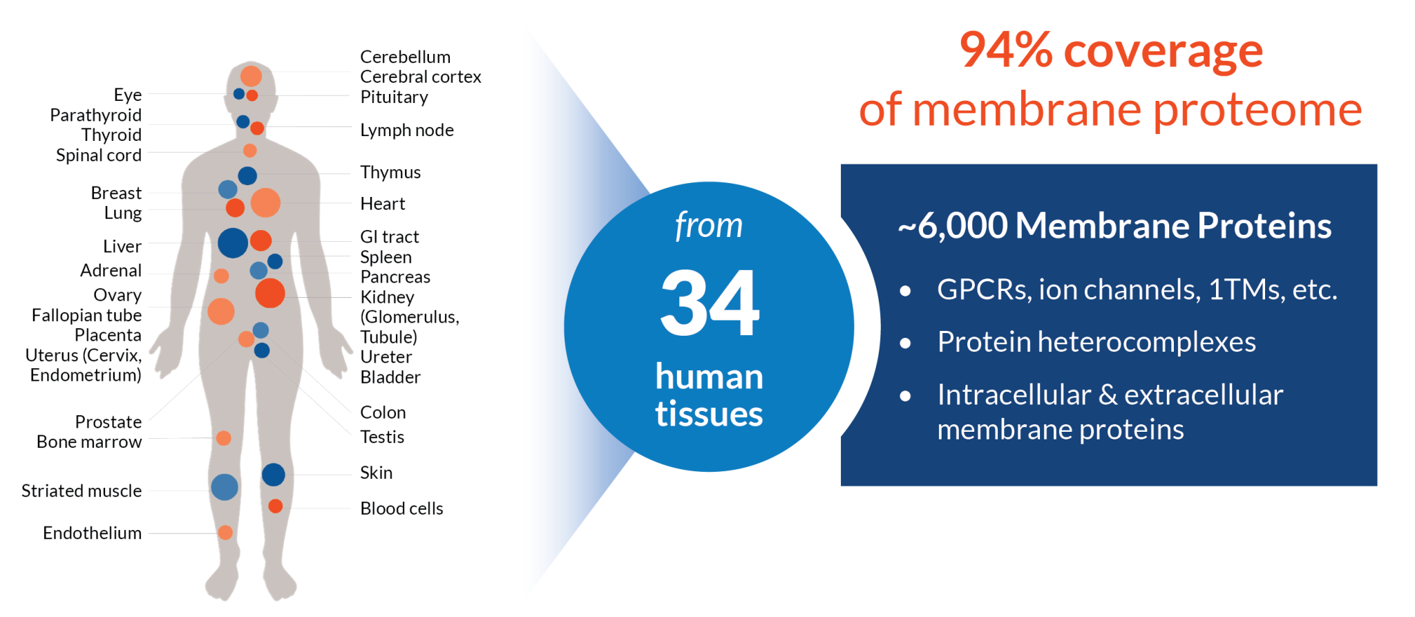
To build the library, our team used a bioinformatics approach to identify all membrane proteins expressed in the 34 specified tissues, drawing on bulk RNA sequencing data, transmembrane topology prediction tools, and mass-spectrometry-derived cell surface protein databases. Full-length canonical isoforms were obtained from UniProt sequences. The result is a library of approximately 6,000 membrane proteins, representing 94% of the human membrane proteome. Importantly, this library spans the full functional range of membrane proteins. The next article in the series will discuss the secreted protein library, which together with the membrane protein library allows for screening across over 7,000 human proteins.
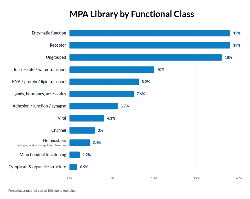
What’s in the library beyond the canonical membrane proteome?
A bioinformatics scan of canonical transmembrane proteins captures the bulk of the membrane proteome, but not all of it. Several categories of proteins that matter for specificity testing could easily be missed by a standard search. The MPA library includes them, along with membrane proteins predicted to be intracellular. Here’s why.
Intracellular membrane proteins. The MPA library deliberately includes membrane proteins thought to localize inside the cell — on the nucleus, Golgi, endoplasmic reticulum, and other organelle membranes. There are two reasons. First, cellular localization is often a prediction, and it’s not fully known which membrane proteins traffic to the cell surface and which do not. Second, even when localization is well-characterized under one set of conditions, it can shift with cell type, activation state, or disease state.
Heterocomplexes. Some membrane proteins only express or fold properly as part of an obligate heteromer; integrins are the canonical example. The MPA library contains approximately 250 heterocomplexes, generally composed of two or three subunits or featuring subunits that participate in multiple complexes. Monomeric subunits of each heterocomplex are also included individually. Because the MPA expresses each protein in a human or other eukaryotic cell, heterocomplexes can also form naturally with the cell’s endogenous proteins.
GPI-linked proteins. Glycosylphosphatidylinositol-linked proteins are anchored to the cell membrane but lack transmembrane domains, which means a standard bioinformatics search targeting canonical transmembrane topology would miss them. Given their importance and surface expression, GPI-linked proteins were curated separately from UniProt. The MPA library includes 120 GPI-linked proteins.
Viral envelope proteins. These are not human proteins and would not be captured by an analysis of the human genome. But they’re important for biotherapeutic development, particularly for antiviral therapies. The MPA library includes 35 envelope proteins from viruses including HIV, dengue, and Ebola.
How well does the MPA represent human variation?
A library can be large without being broadly representative. A specificity testing tool intended to support patient safety needs to address coverage across the dimensions that matter for actual patient populations: protein isoforms, human populations, biological sex, and developmental stages.
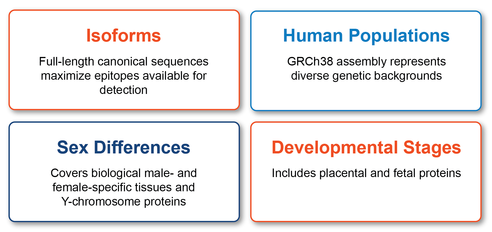
Protein isoforms. The total number of human protein variations, across splice isoforms and post-translational modifications, is beyond the capability of any single in vitro test system. The MPA library is built from canonical isoforms (UniProt), which contain the full target protein sequence; non-canonical isoforms typically delete exons. The library therefore represents the major haplotype and the most complete target sequence for testing binding. Specific isoforms, polymorphisms, or mutations relevant to a particular patient population may need to be evaluated outside of the standard MPA study.
Human population coverage. The MPA is designed for specificity testing across all human populations. The library uses the GRCh38 reference assembly’s canonically defined isoforms. While genetic variation accounts for some differences among humans, gene expression rather than protein sequence is believed to drive most phenotypic diversity across populations (Taylor et al., 2024). Because protein-coding sequences are mostly identical across populations and the MPA overexpresses full-length proteins, the library captures nearly all potential binding targets across human genetic backgrounds. We do note that GRCh38 is skewed toward European and African ethnicities, and the library does not represent disease-specific mutations.
Sex differences. The MPA library is unbiased toward biological sex. Using the Human Protein Atlas, we identified 57 transmembrane proteins with enriched expression in male- or female-specific tissues, nearly all of which are included in the MPA library. We also identified genes on the Y chromosome encoding transmembrane proteins, 10 in total, all of which are included in the MPA library.
Developmental stages. Consistent with the FDA’s TCR panel recommendation, the MPA is designed to represent the adult membrane proteome. However, the library also includes nearly all membrane proteins expressed in the placenta and the fetus, which can be exposed to therapeutics intended for the mother. These inclusions make the MPA better than tissue-based methods for testing binding against developmentally restricted proteins, and eliminate the need to procure fetal tissues.
The breadth of coverage represented in the MPA library is structurally difficult to achieve with tissue-based methods. As discussed earlier in the series, tissue cross-reactivity (TCR) studies draw from a small donor panel, limiting the genetic diversity captured in the study. By working at the protein level and drawing from the reference genome, the MPA library represents the human proteome rather than the biology of a specific set of donors.
Looking ahead
The MPA library is engineered for comprehensive coverage of the membrane proteome — but membrane proteins aren’t the whole specificity story. Secreted proteins can also be off-target binders, with consequences for both safety and pharmacokinetics. In the next article, we’ll look at the Secreted Proteome Library (SPL), which extends the same engineering philosophy to soluble proteins and gives developers a complete picture of their molecule’s interactions across the human proteome.